Analyzing protein function is a key focus in cell biology research. ClontechˇŻs Lentiviral and Retroviral ProteoTuner systems make it possible to
investigate the function of a specific protein of interest directly-by rapidly changing the abundance of the protein itself.
Protein Regulation Mechanism
These systems utilize ligand-dependent destabilization domains (DD) and one of two ligands (Shield1 or Guard1) to reversibly stabilize and destabilize
a DD-tagged protein of interest in a predictable and dose-dependent manner.
- ProteoTuner Shield Systems: Your protein of interest is fused to a DD tag and rapidly stabilized by adding Shield1 ligand to the culture
medium (Figure 2).
- ProteoTuner Guard Systems: Your protein of interest is fused to a DDG tag and rapidly stabilized by adding Guard1 to the culture medium
(Figure 3).
Control Two Proteins Independently
The destabilization domains of the ProteoTuner Shield and Guard Systems are derived from separate protein stabilization technologies.
They are modified from the DDs of FKBP12 (1) and ecDHFR (2) respectively and have been shown to work orthogonally to each other (Figure 1; 2).
The Best Choice for Your Protein
N or C terminal? - Most of the proteins that Clontech has tested show a better destabilization profile when the DD tag is fused to the N-terminus
of the protein of interest (Systems N). Specific DD tag mutants for C-terminal tagging are available as well (Systems C); however they have a slightly
reduced destabilization activity in the absence of the respective ligand.
Tightest control - We believe that for most proteins, ProteoTuner Shield Systems will show greater destabilization, particularly when fused to
the N-terminus of your protein. Unlike the Shield systems, ProteoTuner Guard Systems are only effective at low expression levels. High expression
levels resulting from plasmid transfection lead to high background, due to incomplete destabilization. Although it is possible that plasmid systems
with a weak promoter would work, the best way to limit the expression with ProteoTuner Guard Systems is to control retroviral or lentiviral copy
number and transduce with a low MOI of 0.5 to 5 per cell.
Transmembrane proteins - The ProteoTuner Guard System is likely the best choice for most transmembrane proteins. Iwamoto et al.
(2) showed that fusing DDG (Guard System C) to the C-terminus of CD8Ąá yielded far greater destabilization than could be obtained using
FKB12 technology (Shield System).
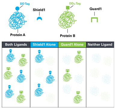
Figure 1. The ProteoTuner Shield and Guard Systems use separate destabilizing domains and stabilizing ligands.
To control two proteins separately but simultaneously, fuse the Shield destabilizing domain (DD) to one protein and the Guard destabilizing domain
(DDG) to the other protein. To stabilize both proteins, add Shield1 and Guard1 simultaneously (left panel). To stabilize just one protein, add either
Shield1 or Guard1 (middle panels). To destabilize both proteins, do not add Shield1 or Guard1 (right panel).
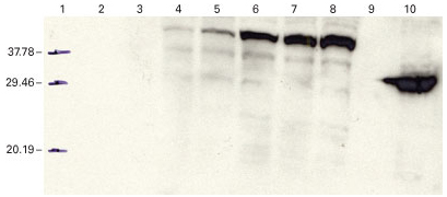
Figure 2. Protein stabilty controlled using the ProteoTuner ShieldSystem. DsRed-Express was cloned into pRetroX-PTuner IRES and
used to infect HeLa cells. The amount of DD-tagged DsRed-Express stabilized by different concentrations of Shield1 was detected via Western
blot using the Living Colors DsRed Polyclonal Antibody. Lane 1: molecular weight marker. Lane 2: 1X loading buffer. Lane 3: untreated HeLa cells
(no virus, no Shield1). Lane 4: HeLa cells infected with the DD-DsRed Express construct; no Shield1. Lanes 5-8: HeLa cells infected with
the DD-DsRed-Express construct and treated with 50, 250, 500, and 1,000 nM Shield1 respectively. Lane 9: 1X loading buffer.
Lane 10: HEK 293 DsRed-Express stable cell line.
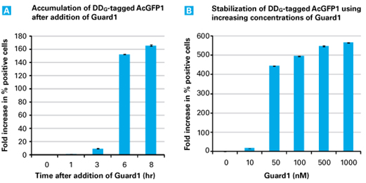
Figure 3. Protein stabilty controlled using the ProteoTuner Guard System. AcGFP1 was cloned into the pLVX-PTuner2 Vector
downstream of the DDG and transfected into packaging cells. Viral particles were harvested and used to infect HeLa cells, following
standard infection procedures using polybrene and a 1 hr spin. 6 hr post-infection, the media was removed and replaced with new media and
cultured for 14 hr. Panel A. The media was removed from the infected HeLa cells and replaced with media containing Guard1 (500 nM).
At the indicated timepoints, cells were collected and analyzed via flow cytometry. Panel B. The media was removed from the infected HeLa cells
and replaced with media containing different concentrations of Guard1 as indicated. 15 hr later, cells were collected and analyzed via flow cytometry.
References
References
1. Banaszynski, L.A. et al. (2006) Cell 126(5):995-1004.
2. Iwamoto, M. et al. (2010) Chemistry & Biology 17: 981-988.
Components
Each system consists of a vector and the destabilization domain's ligand (Shield1 or Guard1)
Items available separately:
-Shield1
-Guard1
-DD Monoclonal Antibody (for use with Shield systems)
Storage Conditions
Store Shield1, Guard1, and all plasmids at -20ˇĆC