- High Efficiency DNA Delivery - 3D cultureНУ ДйОчЧб cellПЁМ Дѕ ГєРК ЙпЧіРЛ АЁДЩЧЯАд Чд
- Easy-to-Use - КАЕЕ УжРћШ АњСЄРЛ УжМвШЧб АэАД ФЃШРћ ЧСЗЮХфФн
- Flexibility - Solid matrix scaffoldПЭ hydrogel 3D culture ЦїИф И№ЕЮПЁ КДЧр АЁДЩ
- Animal Origin Free -ДйОчЧЯАэ ГаРК РћПыРЬ АЁДЩ
0.4 ml TransIT-3D Transfection Reagent & 2 x 12-well alvetex plates
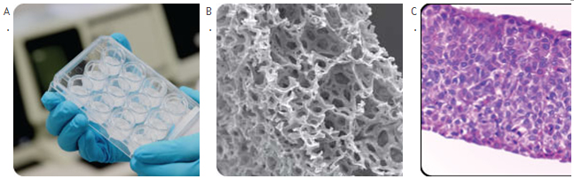
Figure 1. AlvetexЂч Scaffold for 3D Cell Culture. (A) Presentation of alvetex in a 12-well plate format. (B) Alvetex is a highly porous cross-linked polystyrene scaffold, which as been sectioned into 200 Ѕьm thick membranes. (C) Cross section of HaCaT cells grown for 3 days in submerged media conditions and 14 days at the air interface.
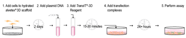
Figure 2. 3D Transient Transfection Protocol. Using the 3D Transfection System, cells are quickly adapted to 3D culture in alvetexЂч 12-well plates. Post-adaptation, the cells are transfected with high efficiency and minimal optimization using the TransITЂч-3D Transfection Reagent.
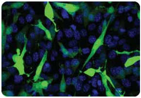
Figure 3. GFP Expression in 3D Culture. NIH3T3 fibroblast cells were seeded at optimized cell density in alvetexЂч 12-well plates and adapted to 3D growth. Forty-eight hours post-adaptation, cells were transfected with TransITЂч-3D combined with a plasmid encoding Green Fluorescent Protein (GFP). Cells were counterstained with the nuclear stain Hoechst 33342 (blue) and visualized via confocal microscopy.
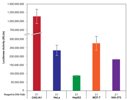
Figure 4. 3D Transfection of Multiple Cell Types. The indicated cell lines were seeded at optimized cell densities in 12-well alvetexЂч 3D plates and adapted to 3D culture conditions for 48 hours. Post-adaptation, cells were transfected with TransITЂч-3D combined with a plasmid encoding firefly luciferase at the reagent-to-DNA ratios indicated. Luciferase activity was measured 24 hours post-transfection using a conventional assay.