- Broad Spectrum DNA Delivery - Achieve high expression in many cell types, including hard to transfect cell lines and primary cells
- Outperforms Competitor Reagents - TransITĒį-2020 demonstrated higher protein yield when compared to FuGENEĒį HD, Lipofectamine™ 2000, and Lipofectamine™ 2000 CD
- Superior Transfection of Insect Cells - Obtain higher expression than other insect cell transfection reagents .
- Animal Origin Free - TransITĒį-2020 provides high performance with maximum compatibility .
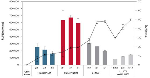
Figure 1. TransITĒį-2020 Reagent Exhibits Higher Expression and Lower Cellular Toxicity Compared to Other
Transfection Reagents. Human umbilical vein endothelial cells (HUVEC) were transfected with a luciferase
expression plasmid using the designated reagents at the reagent-to-DNA ratios indicated beneath each bar.
Transfections were performed in 96-well plates using 0.1 Ĩėg of plasmid DNA per well. Luciferase expression
(bar graph) and lactate dehydrogenase (LDH) levels (line graph) were measured at 24 hours post-transfection.
LDH levels are reported as % cytotoxicity compared to cells alone and were measured using a commercially available
colorimetric assay; all values at or below zero are represented as zero on graph. Error bars represent the standard
deviation of triplicate wells.
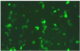
Figure 2. High Performance Plasmid Transfection. Primary Human Small Epithelial Cells (HSAEpic) were transfected
using TransIT-2020 and an EGFP expression plasmid (4:1 reagent-to-DNA ratio). Images were taken 24 hours post-transfection
using a Zeiss axiovert inverted fluorescence microscope.
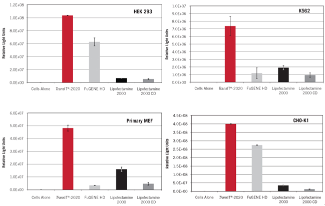
Figure 3. Superior Gene Expression in a Broad Spectrum of Cell Types. Luciferase gene expression was compared in HEK 293,
K562, CHO-K1, and primary MEF cells transfected with a luciferase expression plasmid using TransIT-2020 (Mirus Bio, 3:1 reagent to
DNA ratio), FuGENE HD (Roche, 7:2 reagent to DNA ratio), Lipofectamine™ 2000 (Invitrogen, 5:2 reagent to DNA ratio), and
Lipofectamine™ 2000 CD (Invitrogen, 5:2 reagent-to-DNA ratio). Transfections were performed in 24-well plates using 0.5 Ĩėg of
plasmid DNA per well. The optimal level of each transfection reagent was determined empirically, and all reagents were used according
to manufacturerĄŊs protocol. Cells were harvested at 24 hours post-transfection and assayed for luciferase activity.
Error bars represent the standard deviation of triplicate wells.
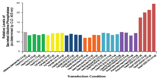
Figure 4. TransITĒį-2020 Reagent Effectively Transfects Drosophila S2 Cells. Cells were transfected with a plasmid construct
expressing a secreted form of the Dscam extracellular domain fused to alkaline phosphatase (AP) in a 24-well plate.
The ratio of transfection reagent to DNA and micrograms of DNA per well is noted beneath each bar.
All products were used according to manufacturersĄŊ protocol. All transfections were performed in serum-free media
for four hours followed by complete media supplementation. An AP enzymatic assay was used to measure the AP levels 24 hours post-transfection.
Data courtesy of Woj Wojtowicz, University of California, Berkeley