Selection Guide: Fluorescent Proteins
Use the list below to choose the best fluorescent protein vector for your experiment based on color/application or name. Click on the name to visit the product page for more information.
| Choose by color: | |||
|
Color |
|||
Cyan Fluorescent Protein
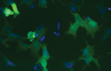
Green Fluorescent Protein
Yellow Fluorescent Protein
Orange Fluorescent Protein
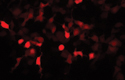
Red Fluorescent Protein
Far-Red Fluorescent Protein
| HcRed1 | 588/618 | 4% | C.T. 41043; Omega XF102-2 |
| mRaspberry | 598/625 | 80% | C.T. 41043 |
| E2-Crimson | 611/646 | 180% | C.T. 41019 |
| mPlum | 590/649 | 25% | C.T. 41043 |
Switchable Fluorescent Proteins
| Choose by application: | |||
|
Application |
|||
Bacterial Expression
 |
We offer a variety of fluorescent protein vectors for bacterial expression. Fluorescent protein expression in these vectors is driven by the lac promoter. |
Cell Capture & Enrichment
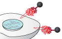 |
CherryPicker Systems use a membrane-targeted red fluorescent protein to identify your cells of interest. These cells can be captured using our specially-formulated magnetic beads and regrown into a more homogenous population in <2 hr. |
Cell Cycle Reporters
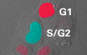 |
Fucci cell cycle reporter vectors deliver fluorescent, ubiquitination-based cell-cycle indicators that allow you to identify cells in various phases of the cell cycle. Fucci vectors contain Cdt1 or Geminin, proteins whose levels fluctuate differently throughout the cell cycle, plus a red or cyan fluorescent tag. |
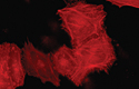
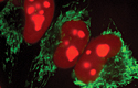
Cell Labeling
 |
Bright colors are ideal for cell labeling and imaging. |
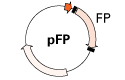
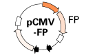
Fusions
 |
Monomeric fluorescent proteins are often ideal for fusions as they tend to be least disruptive to protein function. In many cases, oligomers can also be effective choices. We offer N- & C-terminal vectors. |
FRET
 |
Detecting protein-protein interactions requires a good (high efficiency) FRET pair: a donor with a high quantum yield (QD) and an acceptor with a high Förster Radius (R0). These pairs have been reported to be suitable for FRET in the literature. |
|
In Vivo Imaging
 |
Far red and red fluorescent proteins are preferred for in vivo imaging because they avoid the natural green autofluorescence produced by plants and animals; however, bright green proteins have also been used successfully. |
Kinetic Studies
 |
Destabilized fluorescent proteins, specifically engineered for rapid turnover rates. |
Measuring Proteasome Activity
 |
Our Proteasome Sensor Vector is ideal for image-based assays for compounds with proteasome inhibiting or activating properties. |
MicroRNA Expression
 |
These vectors provide strong miRNA expression and allow you to verify and track that expression with a fluorescent protein marker. The pmR-mCherry and pmR-ZsGreen1 vectors couple your miRNA expression cassette to a bright red or green reporter, for miRNA expression you can see and select. |
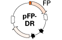
Photoactivation
 |
Photoactivatable fluorescent proteins are particularly useful for determining protein half-life and protein transport pathways, because molecules synthesized after activation are not able to fluoresce. Therefore, what you observe is a snapshot of the protein molecules that were present at the time of activation. |
Photoconversion
 |
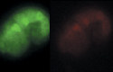
Dendra2 is a photoswitchable (green to red), monomeric fluorescent protein derived from octocoral Dendronephthya sp. Irreversible switching from green to red is activated by UV-violet or blue light. |
Promoter Reporters
 |
Bright fluorescent proteins make excellent reporters. Monitor the activation of different promoters or promoter/enhancer combinations using traditional promoterless reporter constructs or next-generation, on-demand reporters, which use ProteoTuner technology to provide extremely low background and bright signals. |
Stem Cell Applications
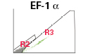 |
Fluorescently labeled stem cells can be used to analyze features and behaviors, and to monitor events including interactions with adjacent cells with precise spatial and temporal resolution. EF-1 alpha promoter vectors are useful in embryonic stem cells, where the CMV promoter has diminished activity. |
Subcellular Labelling
 |
Our subcellular labeling vectors allow you to target a wide range of structures, including actin, cytoskeleton, endosomes, ER, golgi apparatus, mitochondria, nucleus, peroxisomes, and the plasma membrane. |
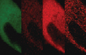
Timed Gene Expression
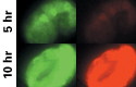 |
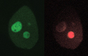
Our pTimer fluorescent protein shifts from green to red over time so you can visualize the time frame of promoter activity. |
Visualizing Gene Expression
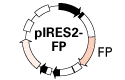 |
IRES (bicistronic) vectors permit your gene of interest and a fluorescent protein to be independently translated from a single RNA transcript. Good for monitoring transfection efficiency or gene expression. |