


- 고객지원
- 업무안내│제품문의
- 전화번호│02-2081-2510
- Email │support@takara.co.kr
- 업무시간안내
- [ 평 일 ] 09 : 00 ~ 18 : 00 │ [ 점심시간 ] 12 : 00 ~ 13 : 00
- 토·일요일, 공휴일은 휴무입니다.
[적용] NDiff 227을 이용한 mouse organoid 제작

Generation of embryonic organoids using NDiff 227 neural differentiation medium
Data kindly provided by Susanne van den Brink, PhD student at Alexander van Oudenaarden laboratory, Hubrecht Institute, Utrecht, Netherlands
- Mouse embryonic stem cell (mESC)로부터 많은 수의 gastruloids (embryonic organoid)를 간단하게 제작하는 방법
- in vitro에서 Mouse 배아 발달 과정의 high-throughput 분석
- 이식 후, mouse 배아와 유사한 80~90% 이상의 긴 형태의 응집체 형성 확인
- 최대 50% 정도의 응집체에서 somite와 같은 구조로 유도됨을 확인
[ Introduction ]
초기의 배아 발달 과정에서 세포 간 상호작용을 통해 작고 균일한 세포 군집을 3차원의 다세포 organism이 되도록 조절한다. 이러한 배아 발달 과정에서 나타나는 오류는 발달 이상으로 이어질 수 있지만, 이런 비정상적인 증상이 언제, 어떻게 유발되는지는 거의 알려진 바가 없다. 마우스와 같은 동물모델을 통해 대부분의 배아 발달 과정에 대한 연구가 진행되어 왔지만, 마우스 배아를 장기적으로 배양하고, 확인하는 것은 기술적인 한계가 있었다. 게다가, 유전자가 조작된 배아를 제작하는 것은 시간이 많이 소요될 뿐 아니라 비효율적이며, 큰 규모의 요구되는 유전자 또는 약물 스크리닝 분석을 위해 마우스 배아를 대량으로 얻는 것이 어렵다는 한계가 있다.
마우스 배아로부터 유래한 mouse embryonic stem cell (mESC) 연구가 발달하면서, 위의 한계를 극복하고 in vitro 상에서 pluripotency를 유지함과 동시에 large-scale의 배아 발달 연구를 가능케했다. 이후로 2차원과 3차원의 mESC 배양 시스템이 개발되었지만, 배아의 복잡한 3D 형태를 정확하게 재현해 내지 못했다. 따라서, 이런 배양 시스템들은 배아의 발달 과정에 관여하는 배아나 배엽 간의 3D 상태에서의 상호작용이나 형태적 재배열, 혹은 배아의 organ 분화 과정을 완벽하게 이해하기 위해서는 사용되기 어려웠다.
이 한계를 극복하기 위해, 네덜란드 Hubrecht Institute의 Susanne van den Brink와 동료들은 mESC를 이용해 organoid 구조의 3D 배양법을 개발하였다. Gastruloids 구조는 삼배엽이나 body axis와 같이 배아 이식 후에 보이는 현상들을 보였다 (van den Brink et al. 2014; Turner et al. 2017; Beccari et al. 2018; van den Brink et al. 2020). 이러한 구조를 제작해내기 위해, 연구자들은 Takara Bio의 NDiff 227 배양 배지를 이용해 초기 배아 단계와 유사하도록 mESC를 효율적으로 응집하고 재현성 높게 분화 시키고자 했다. 이후에 Wnt agonist를 적용하여, 이 응집체는 점차 길어지면서 머리와 꼬리를 이루는 전-후 body axis에 따라 적절한 위치에 구조체가 형성됨을 확인하였다. 이와 같이 배아 발달 과정을 재현하는 organoid 구조는 gastruloids라고 불리며, in vitro 상에서 마우스 배아 발달을 연구하는 데 사용될 수 있다.
[ Results ]
NDiff 227는 defined, serum-free 배지로, mESC를 adherent monoculture하여 neural lineage로 분화 시키기 위해 개발되었다 (Ying et al. 2003). 그러나 연구자들은 NDiff 227 배지가 mESC로부터 3D embryo-like organoid structure (gastruloids)를 높은 재현성으로 형성하는 데에도 효과적이며, 이를 in vitro 내에서 배아 발달에 대한 연구를 high-throughput 으로 적용할 수 있음을 새롭게 확인하였다.
이 프로토콜에서는 부착성이 낮은 U-bottom 96 well plate의 각 well에 300개 가량의 mESC를 분주하고, NDiff 227 배지에서 배양 및 응집시켰다 (Figure 1). 분주 후 2일 동안에는 세포는 세포끼리 응집되어 하나의 구형을 이루며, 각 well의 바닥에 가라앉게 된다. 이후 배양 3일차에 Wnt-agonist Chiron (CHIR99021)을 처리하여 24시간 배양하게 되는데, 처음 분주한지 4~5일차 즈음에 구형으로 대칭을 이루었던 형태가 길쭉한 형태로 변화한다. 이 구조는 시간이 지남에 따라 점차 위아래로 더 길어지는 형태를 보였으며 (van den Brick et al. 2014), 마우스 배아에서 형성하는 세포 유형이 생성되고 머리와 꼬리를 이루는 전-후 body axis에 따라 각 정확한 위치에 배치되는 것이 확인되었다 (van den Brick et al. 2020). 이렇게 생성된 구조체는 신체를 구성하는 삼배엽으로의 분화 또한 확인되었다.
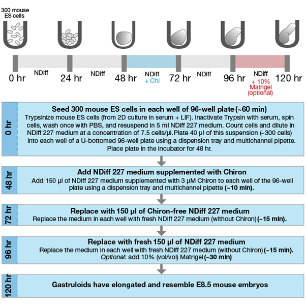
그림 1. mESC로부터 gastruloids가 형성되는 과정
Gastruloids are produced by plating 300 mouse ES cells per well in NDiff 227 medium in a U-bottomed 96-well plate. After seeding, the plates are placed in the incubator for 48 hr. During the incubation, the cells sink to the bottom of the well and stick together to form a round aggregate. At 48 hr, NDiff 227 medium is supplemented with the Wnt-agonist Chiron (Chi), which induces a symmetry-breaking event. At 72 hr, the Chiron-supplemented NDiff 227 medium is replaced with plain NDiff 227 medium. Between 96 and 120 hr after aggregation, the aggregates elongate and form an embryonic organoid that contains all three germ layers and all three body axes. Optional: adding 10% Matrigel to gastruloids at 96 hr after aggregation induces the formation of somite-like structures and results in gastruloids that resemble mouse embryos more closely. Approximate hands-on time per 96-well plate for each step is indicated within brackets.
2014년에 van den Brink et al. 문헌에서 처음으로 소개한 gastruloids 제작 방법은 길쭉한 형태의 구조물은 형성하였으나 마우스 배아에서 보이는 발달과정의 양상을 모두 확인하기에는 충분치 않았다. 특히 마우스 배아와 달리 gastruloids로부터 somite (배아 뒤쪽에 줄지어 있는 조직 블록으로, 척추와 갈비뼈와 같은 골격을 형성함)가 생성되지 않는 다는 것이 가장 큰 한계였다. 그러나 2020년도에 발표한 문헌에서 연구자들은 배양 96시간 후 낮은 비율의 Matrigel을 응집된 세포 구조에 첨가함으로써 somite와 유사한 조직을 유도할 수 있음을 발견했다. Matrigel의 추가로, 마우스 배아를 보다 정확하게 재현한 gastruloids를 생성할 수 있게 되었으며 (그림 2), NDiff 227 배지를 이용해 in vitro 상에서 많은 수의 gastruloids를 효율적으로 얻을 수 있음을 보여준다.
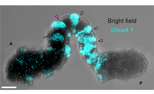
그림 2. Multi-photon microscopy로 촬영한 In vitro gastruloid의 somite 유사 구조
A mouse gastruloid was embedded in 10% Matrigel at 96 hr, later stained for Uncx4.1 (which marks the posterior half of somites), and then visualized at 120 hr using hybridization chain reaction (HCR) imaging. Pink arrowheads point at the boundaries between the individual somites. A: anterior (head); P: posterior (tail). Scale bar = 100 μm. Image credit: Vincent van Batenburg.
[ Conclusions ]
Van Oudenaarden 연구실의 연구자들은 다카라바이오의 neural 분화를 위한 NDiff 227 배지가 in vitro 상에서 mESC로부터 embryonic organoid (gastruloid)를 효율적이고 재현성 있게 제작할 수 있어, 배아 발달과정의 연구를 대량으로 진행하는 데 유용함을 증명하였다. Gastruloid의 생산은 기존에 사용되던 방법과 비교했을 때 뚜렷한 장점을 보였다; (1) 마우스 배아보다 유전정보를 교정하기에 수월함 (2) Body axis 형성과 같이 기존의 in vitro 배양 시스템에서는 어려웠던 구조를 형성하면서, 이에 대한 연구를 가능케 함 (3) 보다 손쉽게 대량의 gastruloid를 제작할 수 있어 약물 스크리닝과 같은 large-scale의 실험에도 적용 가능함
Gastruloid를 제작하는 프로토콜은 일반적으로 사용되는 배양 장비만을 이용하면서 최소한의 세포 배양 경험만이 있더라도 빠르고 간단하게 적용할 수 있으며, 다른 실험실에서 이 in vitro 시스템을 이용하더라도 각 batch별 실험 오차가 적어 재현성이 높은 결과를 얻을 수 있다.
[ Methods ]
mESC는 37ºC; 5% CO2 incubator 내에서 serum과 leukemia inhibitory factor (LIF) 조건으로 배양되어 진다. 세포 응집을 진행하기 전에, 각 세포들은 trypsin으로 well에서 떼어낸 후, PBS로 washing한 후 Takara Bio.의 NDiff 227 배지에서 resuspension 된다. 각각의 300개 정도의 세포를 U-bottom 96 well에 분주하고, 40 ㎕의 NDiff 227 을 첨가하여 48시간동안 배양한다. 이 기간 동안 세포는 하나의 구형 응집체를 형성하여 각 well 바닥에 가라 앉게 된다. 배양 48시간 후, 3 uM Chiron (CHIR99021)을 포함하는 150 ul의 NDiff 227 배지를 multichannel pipette을 이용해 첨가하고, 24시간 동안 추가로 배양한다. 이후 Chiron을 포함하는 NDiff 227 배양 배지를 well에서 제거하고, Chiron을 포함하지 않는 새로운 NDiff 227 배지 150 ㎕로 교체하여 24시간 동안 추가로 배양한다. 배양 후 총 96시간이 지난 후에는 배지는 마지막으로 새로운 NDiff 227 배지 150 ㎕로 교체하게 되고, 배양 후 총 120시간이 지난 후에는 80-90 % 정도의 응집체에서 배아와 유사한 길쭉한 형태를 확인할 수 있다.
선택사항) 응집 96시간 후에 NDiff 227 배지에 10% Matrigel을 첨가하게 되면, 최대 50% 정도의 응집체에서 somite와 유사한 구조의 형성을 유도할 수 있다. Somite와 같은 구조체를 포함/포함하지 않는 gastruloid의 제작 프로토콜은 아래의 논문에서 확인할 수 있다. 이전 버전의 프로토콜은 비디오 형태로 확인할 수 있다.
- [METHOD ARTICLE] Generating gastruloids with somite-like structures from mouse embryonic stem cells (클릭)
- [Video] Generation of Aggregates of Mouse Embryonic Stem Cells that Show Symmetry Breaking, Polarization and Emergent Collective Behaviour In Vitro (클릭)
miPSC를 이용하는 경우에도, NDiff 227 배지는 응집체 형성에 적용 가능하다는 결과가 확인되며, 보다 자세한 miPSC 기반의 mouse gastruloid 제작 프로토콜은 아래에서 확인할 수 있다.
- [METHOD ARTICLE] Generating Gastruloids from Mouse Embryonic Stem Cells (클릭)
[ References ]
- Beccari, L. et al. Multi-axial self-organization properties of mouse embryonic stem cells into gastruloids. Nat. Lett. 562, 272-276 (2018).
- van den Brink, S. C. et al. Single-cell and spatial transcriptomics reveal somitogenesis in gastruloids. Nature 582, 405-409 (2020).
- van den Brink, S. C. et al. Symmetry breaking, germ layer specification and axial organisation in aggregates of mouse embryonic stem cells. Development 141, 4231-42 (2014).
- Turner, D. A. et al. Anteroposterior polarity and elongation in the absence of extra-embryonic tissues and of spatially localised signalling in gastruloids: mammalian embryonic organoids. Development 144, 3894-3906 (2017).
- Ying, Q. et al. Conversion of embryonic stem cells into neuroectodermal precursors in adherent monoculture. Nat. Biotech. 21, 183-86 (2003).
[원문] Generation of embryonic organoids using NDiff 227 neural differentiation medium