Deconvolution, segmentation 또는 복잡한 알고리즘 없이 분석하여 진정한 true single cell 분석 가능
Nuclei에 tagging된 spatial barcode로 single cell data의 높은 품질과 민감도 유지
기존 single cell NGS workflow에 Trekker® kit를 추가하여 true single cell spatial data 확보
Trekker® Single-Cell Spatial Mapping Kit Workflow
기존 single cell NGS 분석의 upstream으로 Trekker®를 추가하면 전용 장비 없이 간단하게 spatial data 추가 가능
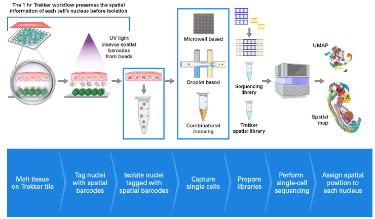
그림 1. Trekker® 기술의 핵심은 spatial barcode가 결합된 bead가 단층으로 구성된 tile에 있습니다.
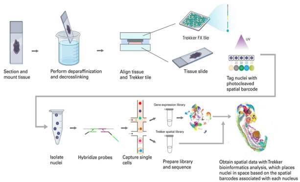
25 μm 두께의 동결절편 (fresh frozen tissue)을 Trekker® tile 위에 올린 후 UV를 조사하면, bead에 결합되어 있던 spatial barcode가 절단되어 주변의 핵 (nuclei)에 tagging 됩니다. 그 후 조직을 Trekker® tile에서 분리하여 핵을 회수한 뒤, single-nucleus RNA sequencing (snRNA-seq)을 수행합니다. 이 과정에서 spatial barcode는 RNA와 함께 증폭되며, 최종적으로 single-cell의 유전자 발현 (gene expression) library와 spatial library의 두 가지 library가 생성됩니다. 최종 데이터는 전용 분석 파이프라인을 통해 각 핵이 보유한 spatial barcode를 기반으로 조직 내 원래 위치를 mapping하여, true single-cell 수준의 spatial transcriptomics 데이터를 생성할 수 있습니다. Trekker® 프로토콜은 기존 snRNA-seq 워크플로우 전 단계에 약 1시간의 과정만 추가하면 적용할 수 있습니다.